
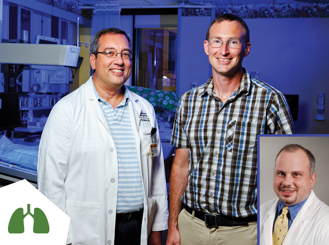
Intelligent oxygen control for NICU patients
Principal Investigators
Roger Fales, PhD
Department of Mechanical and Aerospace Engineering
John Pardalos, MD
Department of Child Health
Ramak Amjad, MD
Department of Child Health
Each year, many of the more than 28,000 babies born with low birth weight and 41,000 neonatal intensive care unit (NICU) patients require respiratory support because of immature lung development and other problems associated with prematurity. Respiratory support might last for weeks, or even months, during a NICU stay. Infants in the NICU have widely varying responses to oxygen control, so they require continuous monitoring and manual adjustment of oxygen to avoid apnea and oxygen desaturation. If blood-oxygen saturation is too high and fluctuates frequently, infants can suffer from retinopathy of prematurity (ROP), the leading cause of blindness and visual impairment in premature infants.
Currently, pulse oximeter alarms notify medical staff if an infant is outside of the desired levels of oxygen saturation, and staff members must make manual adjustments. As multicenter data has shown that premature infants spend only about half of the time within the prescribed range of oxygen saturation during manual care, the principal investigators have developed a device that automatically varies oxygen and/or airflow as needed. The device uses feedback from multiple sensor measurements, such as blood-oxygen saturation, heart rate and respiratory rate, to increase the amount of time babies spend in the desired range of oxygen saturation. The device is “intelligent” because a control algorithm allows it to learn and adapt over time so that blood-oxygen saturation is controlled more reliably and accurately even with wide variations in patient condition. The researchers have completed simulation studies and are preparing for human testing.
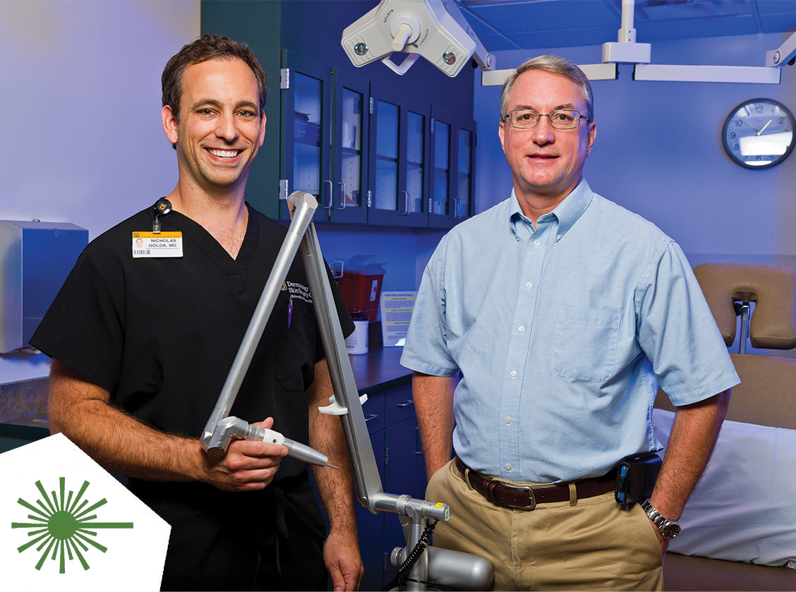
Safer laser handpiece for dermatology treatments
Principal Investigators
Randy Curry, PhD
Department of Electrical and Computer Engineering
Nicholas Golda, MD
Department of Dermatology
With more than 7,800 practicing laser dermatology clinics and an additional 2,100 medical spas, lasers are becoming more common as minimally invasive dermatological tools. Current handpieces have fundamental design flaws that present extreme ocular hazards to the practitioner and patient, fail to account for the negative tissue effects that result from laser illumination of skin and are not ergonomically designed with the practitioner in mind. Because there are no alternatives, these flaws are accepted, and this limits treatment efficiency and represents the potential for serious injury.
The principal investigators are developing a laser device accessory that will make procedures safer and more effective and place less strain on practitioners. Their ergonomically designed product functions by emitting laser light directly into the skin through physical contact. This eliminates the risk of serious, irreparable ocular injury that can result from accidental exposure to even just a reflection of the laser beam. Their design also mitigates the undesirable photo-thermal effects at the tissue surface, including carbonization and vaporization followed by layers of coagulation and edema that currently limits laser exposure energies and treatment times and reduces procedural efficacy. Although some newer methods attempt to utilize contact cooling techniques, most techniques are fundamentally incompatible with contemporary light delivery methods.
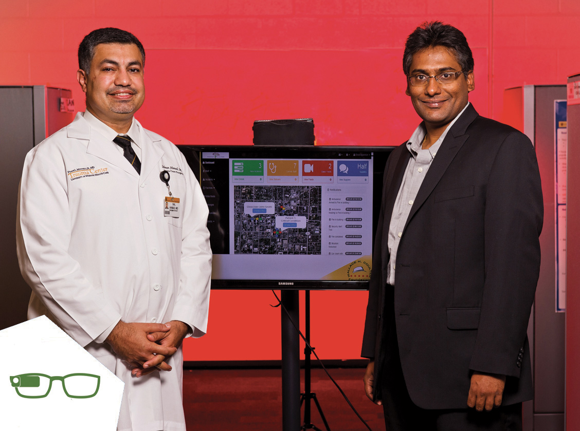
Panacea's Cloud: Augmented reality system for mass casualty disaster triage and coordination
Principal Investigators
Prasad Calyam, PhD
Department of Computer Science
Salman Ahmad, MD
Department of Surgery
Communication in a mass casualty disaster scene is limited and difficult because of the absence of necessary infrastructure. As time and situational awareness are critical, the lack of an integrated technology product that allows a medical director or incident commander to evaluate the scene and strategically delegate triage responsibility leads to misdirected and delayed triage of critically injured patients, potentially causing loss of lives. According to IBISWorld 2014-Report, medical responders are a major portion of the disaster and emergency relief services market, which covers more than 3,700 businesses and is valued at $8 billion.
To meet the need for a robust, portable system that enables audiovisual communications in disaster situations, the principal investigators have developed Panacea’s Cloud, an application that provides augmented reality benefits through integration of a standardized Incident Command System (ICS) with the Internet of Things (IoT). Panacea’s Cloud allows multiple users to communicate with each other using a battery-powered hands-free wireless heads-up display (HUD) unit (e.g. Google Glass, Recon) over a stand-alone battery-powered wireless network. The use of this system, coupled with virtual beacons and QR code cards deployed around the scene, creates a resilient mesh network independent of existing infrastructure that might have been compromised in a disaster. The software features a simple video chat application and a web-based customizable Intelligent Dashboard for response team communication and prioritized coordination. Panacea’s Cloud has been evaluated in a simulated mass casualty environment to show its ease of use and effectiveness in increasing triage care levels. Testing in ambulances is planned.
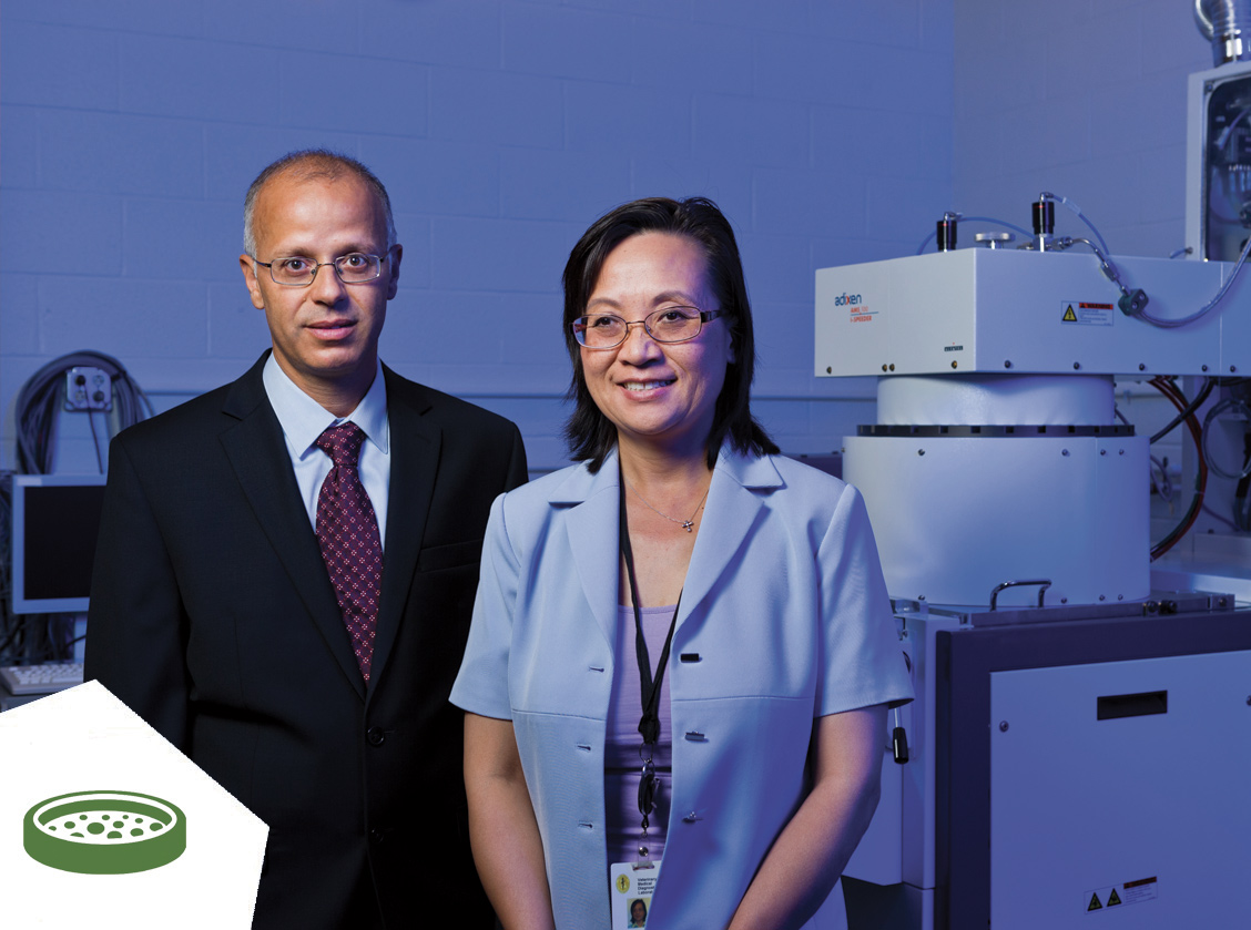
Germ Sensor system for rapid detection of salmonella and other pathogens
Principal Investigators
Mahmoud Almasri, PhD
Department of Electrical and Computer Engineering
Shuping Zhang, PhD, DACVM
Department of Veterinary Pathobiology
Salmonella is a significant food-borne pathogen that causes millions of infections annually in the U.S. Early detection of salmonella in clinical samples is necessary for prompt treatment and control of the infection. Timely detection and removal of contaminated food products — such as fruits, vegetables, water supplies, meats and poultry — can ensure the safety of consumers and help prevent food-borne outbreaks of salmonellosis, which can lead to large financial losses because of medical costs and product recalls. The traditional methods for pathogen detection have relied on microbiological culture and colony counting. These methods are time-consuming (three to five days to identify the contaminant), require trained personnel and cannot be used in the field, making them unsuitable for timely assessment. Numerous groups have tried to develop techniques to reduce pathogen detection time, however, high cost, long reaction times and/or lack of sensitivity and specificity have precluded their implementation for on-site detection.
The principal investigators have designed, modeled, fabricated and characterized an impedance-based MEMS biosensor prototype that will enable rapid detection of very low levels of salmonella in food products. The high sensitivity of their device is the result of a novel design that utilizes microchannels with varying diameters to concentrate the bacteria. The detection region uses an electrode functionalized with anti-salmonella antibodies to selectively detect salmonella.
DR sensor for early detection of diabetic retinopathy
Principal Investigators
Raghuraman Kannan, PhD
Department of Bioengineering
Dean Hainsworth, MD
Department of Ophthalmology
One of the major microangiopathic eye complications that arises in both Type 1 and Type 2 diabetic patients is diabetic retinopathy (DR). It is the leading cause of blindness in adults, with 4.2 million cases reported in the U.S. alone. Of those, 665,000 are vision threatening. Every year, DR results in approximately 24,000 new cases of blindness in adults ages 25 to 74. At any stage of the disease, macular edema can cause severe damage to vision and at the most advanced stage, permanent vision loss can occur. Even though the complications are overwhelming, patients in advanced stages of DR are typically asymptomatic. As no cure exists for DR, vision loss is preventable only by timely detection and treatment. Screening for DR is currently performed by ophthalmologists and optometrists during annual eye exams. Unfortunately, these eye exams are expensive and time-consuming, and annual checkups alone are not sufficient for early detection of DR. In fact, patients can lose up to 60 percent of their vision in between annual checkups. Diabetic patients who do not have regular eye exams are at even greater risks of preventable vision loss.
In order to meet the need for a DR screening test that is inexpensive and suitable for use in primary care settings, the investigators have developed the DR Sensor, a colorimetric nanosensor that detects levels of a urine biomarker indicative of diabetic retinopathy. Current methods for measuring this biomarker require expensive laboratory instruments and lengthy procedure times. The DR Sensor utilizes microfluidic channels impregnated in paper, making the sensing element inexpensive, reliable and disposable. Ultimately, the DR sensor will be coupled with image processing via a simple app that integrates with devices in clinical labs for real-time diagnosis of DR.