Currently, doctors have to throw away more than 80 percent of donated tissue used for joint replacements because the tissue does not survive long enough to be transplanted. Now, following a recent study, University of Missouri School of Medicine researchers have developed a new technology that more than doubles the life of the tissue. This new technology was able to preserve tissue quality at the required level in all of the donated tissues studied, the researchers found.
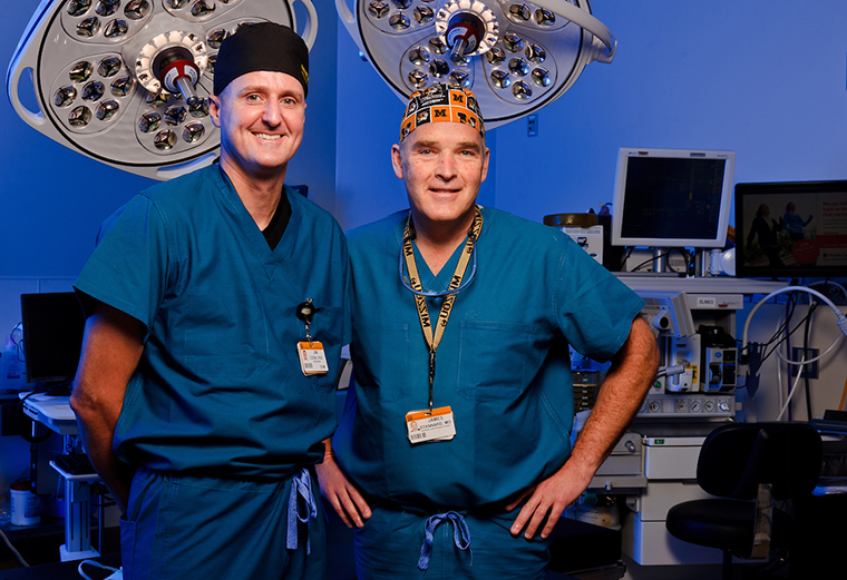
“It’s a game-changer,” said James Stannard, co-author of the study and J. Vernon Luck Sr. Distinguished Professor of Orthopaedic Surgery at the MU School of Medicine. “The benefit to patients is that more graft material will be available and it will be of better quality. This will allow us as surgeons to provide a more natural joint repair option for our patients.”
The technology, called the Missouri Osteochondral Allograft Preservation System, or MOPS, more than doubles the storage life of bone and cartilage grafts from organ donors compared to the current preservation method used by tissue banks.
In traditional preservation methods, donated tissues are stored within a medical-grade refrigeration unit in sealed bags filled with a standard preservation solution. MOPS utilizes a newly developed preservation solution and special containers designed by the MU research team that allows the tissues to be stored at room temperature.
In the study, clinical outcomes of the standard preservation approach and the new MOPS technology were assessed. Researchers found that by using MOPS, the storage time for donor tissue could be extended to at least 60 days, versus the current storage time of approximately 28 days.
“Time is a serious factor when it comes to utilizing donated tissue for joint repairs,” said study co-author James Cook, director of MU’s Comparative Orthopaedic Laboratory and the Missouri Orthopaedic Institute’s Division of Research. “With the traditional preservation approach, we only have about 28 days after obtaining the grafts from organ donors before the tissues are no longer useful for implantation into patients. Most of this 28-day window of time is used for testing the tissues to ensure they are safe for use. This decreases the opportunity to identify an appropriate recipient, schedule surgery and get the graft to the surgeon for implantation.”
Stannard, who also serves as chair of MU’s Department of Orthopaedic Surgery and medical director of the Missouri Orthopaedic Institute, said that patients with metal and plastic implants often are forced to give up many of the activities they previously enjoyed in order to extend the life of their new mechanical joints.
“For patients with joint problems caused by degenerative conditions, metal and plastic implants are still a very good option,” Stannard said. “When the end of a bone that forms a joint is destroyed over time, the damage is often too extensive to use tissue grafts. However, for patients who experience trauma to a joint that was otherwise healthy before the injury, previous activity levels needn’t be drastically altered if we can replace the damaged area with living tissue.”
Donor tissue grafts have been used for many years as a way to fill in damaged areas of a joint, as an alternative to removing bone and implanting metal and plastic components. The body accepts bone and cartilage grafts without the need for anti-rejection drugs, and the donor tissue becomes part of the joint. However, the method of preserving the grafts themselves has limited the amounts of quality donor tissue available to surgeons.
Additionally, because of testing requirements and logistics, only about 20 percent of grafts are ultimately useable because not enough live tissue cells remain in them after 28 days. In contrast, the study found that the MOPS preservation system resulted in a 100 percent rate of usable tissue grafts at 60 days after procurement.
Cook, who also serves as the William and Kathryn Allen Distinguished Professor in Orthopaedic Surgery at the MU School of Medicine, leads the research team that developed the new bone-and-cartilage-preservation technology to overcome these challenges.
“With our new preservation technique, we can offer more patients a repair that allows their joints to respond to daily activities like they did when the joints were healthy,” Cook said. “Like a normal joint, the implanted tissue can renew itself, resulting in decreased physical limitations to the patient.”
The study, “A Novel System Improves Preservation of Osteochondral Allografts” was recently published in Clinical Orthopaedics and Related Research, a publication of the Association of Bone and Joint Surgeons. A previous study concerning the development of the preservation system was published in the Journal of Knee Surgery in 2012.


