BioJoint Flex: A Simple Solution for Stiff Knees
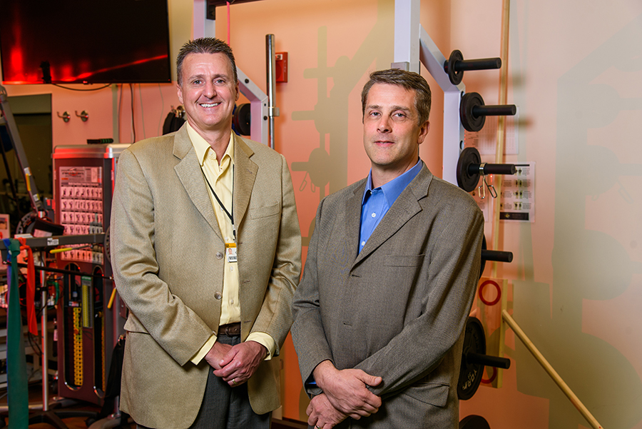
Principal Investigators
Trent Guess, PhD
Department of Physical Therapy and Orthopaedic Surgery
James Cook, DVM, PhD
Department of Orthopaedic Surgery
More than 1 million knee surgeries are done in the United States each year, and an even larger number of patients with knee problems are managed non-operatively. While improved range of motion is strongly linked to improved outcomes, a common limitation for all these patients is loss of knee motion. Existing methods for improving knee range of motion can be painful, costly or ineffective if not done in a controlled and measured manner. The BioJoint Flex is a non-invasive device that assists in controlled, supported and measured knee flexion exercises. This device is specifically designed to safely and effectively improve knee flexion and overall knee range of motion as part of a non-operative or pre/post-operative rehabilitation program in physical therapy clinics, facilities and homes. With the BioJoint Flex, patients can reduce the need for repeat surgery, treat refractory loss of motion (arthrofibrosis) and improve their functional outcomes.

Mizzou Knee Arthrometer Testing System (MKATS): An Easy-to-Use Tool for Accurate Screening, Diagnosis and Treatment Monitoring for Knee Ligament Injuries
Principal Investigators
Trent Guess, PhD
Department of Physical Therapy and Orthopaedic Surgery
James Cook, DVM, PhD
Department of Orthopaedic Surgery
Knee injuries are common, with more than 750,000 ligament injuries and 500,000 anterior cruciate ligament ruptures occurring each year in the United States. When a ligament is torn, knee stability and function become abnormal. Precisely determining the direction and magnitude of the instability (laxity) is vitally important for making a complete and accurate injury diagnosis, determining optimal treatment options and monitoring recovery to determine when it is safe to return to daily activities. While devices that measure knee laxity have been commercially available for decades, the Mizzou Knee Arthrometer Testing System (MKATS) can potentially address limitations of existing devices by delivering cost-effective quantitative measurement of knee laxity. That would be valuable to orthopaedic surgeons, physical therapists and athletic trainers. Additionally, knee laxity is a known risk factor for ACL injury. Assessing this risk with the MKATS would help millions of amateur, collegiate and professional athletes by providing critical decision-making information to physical therapy clinics and athletic training facilities.

OPT-Enhanced Colposcopy: 3D Detection of Precancerous and Cancerous Lesions for Image-Guided Biopsy
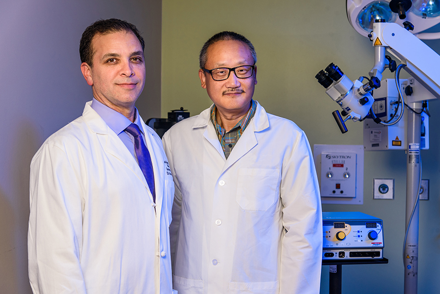
Principal Investigators
Gary Yao, PhD
Department of Bioengineering
Mark Hunter, MD
Department of Gynecologic Oncology
Each year, approximately 2 million women in the United States are referred for colposcopy after positive Pap smear/HRV screening results, and over a half-million women worldwide are diagnosed with cervical cancer. Fortunately, when correctly identified using colposcopy, precancerous lesions (cervical intraepithelial neoplasia or “CIN”) are fully treatable. However, current video-based colposcopy is subjective and requires tissue destruction. Because tissue samples must be sent to the pathology lab for analysis, it has a long lead time for diagnosis. The proposed OPT-Enhanced Colposcopy is a new imaging system based on optical polarization tractography (OPT) technology that makes it possible to accurately and objectively identify and stage CIN, as well as invasive cancerous lesions, during the colposcopy procedure. OPT enables precise 3D imaging of subsurface cellular morphological changes in a living organism. These changes are histology‐comparable markers of precancerous lesions and invasive cancer. OPT-Enhanced Colposcopy could potentially allow clinicians to precisely detect lesions, perform image‐guided therapy and verify treatment outcomes all in a single colposcopy session.

Tongue Twister: Mobile Health App for Early Detection and Monitoring of Tongue Dysfunction Caused by Neurological Disorders
Principal Investigators
Teresa Lever, PhD
Department of Otolaryngology
Mili Kuruvilla-Dugdale, PhD
Department of Communication Science and Disorders
Filiz Bunyak, PhD
Department of Computer Science
Yunxin Zhao, PhD
Department of Computer Science
The tongue is one of the strongest muscles in the body and is essential for normal speech and swallowing function. Therefore, tongue dysfunction caused by neurological disorders such as stroke, Parkinson’s disease, ALS and multiple sclerosis can result in devastating speech and swallowing impairments. In the United States, approximately 15 million people are living with speech and swallowing impairments that profoundly impede quality of life and cost over $30 billion annually in medical care and lost work productivity. Current diagnostic methods to detect and monitor tongue dysfunction are subjective and therefore prone to inaccuracy, which risks misdiagnosis and inappropriate treatment. Tongue Twister provides a low-cost, easy-to-use software solution that transforms widely used tongue performance tasks into real-time objective data, thus enabling earlier and more accurate detection and treatment of tongue dysfunction and monitoring of treatment outcomes. This is especially important for progressive neurological disorders, for which earlier intervention is associated with improved quality of life and survival. Scalability beyond clinical settings includes detection of sports concussions during athletic events and detection of fatigue and substance abuse by law enforcement and safety regulatory agencies.


