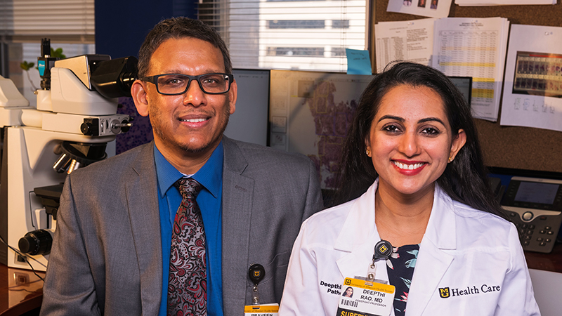
Raghuraman Kannan, PhD  Department of Bioengineering  Gerald Arthur, MD  Department of Pathology and Anatomical Sciences

A novel nanoplatform for accurate detection of biomarkers in tumor tissues

Principal Investigators

Raghuraman Kannan, PhD
Department of Bioengineering

Gerald Arthur, MD
Department of Pathology and Anatomical Sciences


Patient response rates for many targeted cancer therapies are directly dependent on the overexpression of specific molecular targets in tumor tissues. Thus, "companion" diagnostics that can precisely identify and quantify targets such as ligand receptors have become important for the success of targeted therapies. The principal investigators have developed a gold nanorod based histochemistry (GNR-HC) platform that they have proven can identify and quantify a variety of target biomarkers in tumor tissues in ways superior to existing methods.

The first target validated was Epidermal Growth Factor Receptor (EGFR), a biomarker expressed in multiple types of cancer including colorectal adenocarcinoma, non-small cell lung carcinoma (NSCLC), head and neck carcinoma, and glioblastoma.

The second validated biomarker was the cMET protein, which plays a role in progression of NSCLC, colorectal cancer, breast cancer, and gastrointestinal tumors.

The heterogeneous nature of cancer necessitates the identification and quantitation of major/driver mutations in each patient so that a "personalized" treatment plan can be constructed. The principal investigators will now use their platform to develop an in situ hybridization assay for detection of EGFR mutations that have been shown to play an important role in the pathogenesis of NSCLC through the enhancement of tyrosine kinase activity (T790M and L858R). The combined information on protein targets and point mutations will provide the comprehensive data needed to devise a patient-specific treatment plan for NSCLC patients.


A. Sherif El-Gizawy, PhD  Department of Mechanical and Aerospace Engineering  Raja Gopaldas, MD  Department of Surgery

An embolic protection device to aid in transcatheter aortic-valve implantation and prevent neurological dysfunction

Principal Investigators

A. Sherif El-Gizawy, PhD
Department of Mechanical and Aerospace Engineering

Raja Gopaldas, MD
Department of Surgery


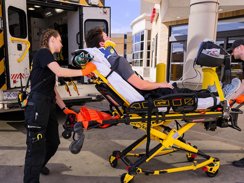
There are approximately 250,000 aortic valve replacement procedures globally per year, with 90,000 of the procedures being done in the United States. Open heart surgery is the current standard of care for aortic valve replacement, but 40% of patients cannot tolerate open heart surgery due to underlying health related issues or age. Within the last two years, the minimally invasive TAVI procedure has become available to these high risk patients. However, 22% of TAVI recipients manifest acute cerebral diffusion abnormalities or intra-operative strokes.

In this project, the PIs have developed a device that creates a protective barrier with-in the aorta preventing embolic debris from entering brain vessels. This technology has been licensed to Cardioptimus. Active fundraising is proceeding to continue development of the


Matthew Smith, MD  Department of Orthopaedic Surgery  Ferris Pfeiffer, PhD  Department of Orthopaedic Surgery and Department of Bioengineering

A bone-tendon allograft system optimizing tissue healing and biomechanical strength for human rotator cuff repair

Principal Investigators

Matthew Smith, MD
Department of Orthopaedic Surgery

Ferris Pfeiffer, PhD
Department of Orthopaedic Surgery and Department of Bioengineering


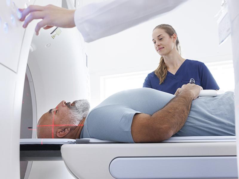
Approximately 600,000 rotator cuff repair procedures are done per year in the United States of which, massive rotator cuff tears account for nearly 30%. The current standard of care for rotator cuff repair is tendon to bone suture repair. However, during massive tears, the resulting tendon quality is not sufficient to allow repair. Many factors are associated with poor outcomes, including tendon retraction, muscle atrophy and fatty degradation, poor tissue quality and difficulties to suture tendon fibers. A failure rate of up to 94% is seen in current massive tendon repairs.

The PIs have developed a system which provides and bone tendon allograft to be deployed arthroscopically to repair the tear. The system prepares a recipient channel to interface with a novel allograft design that optimizes tissue healing and integration as well as biomechanical strength.