The goal of the nuclear cardiology rotation is to train all fellows in the fundamentals of nuclear cardiology for application in the clinical management of cardiovascular patients.
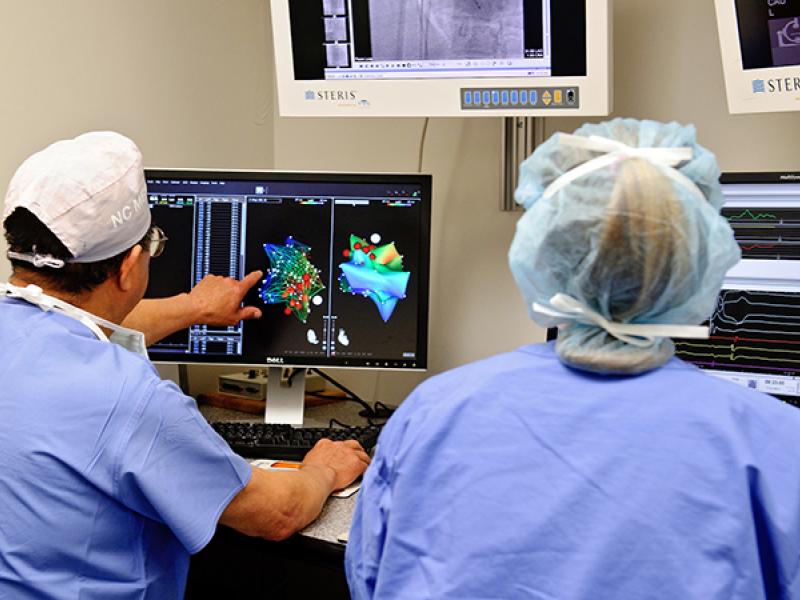
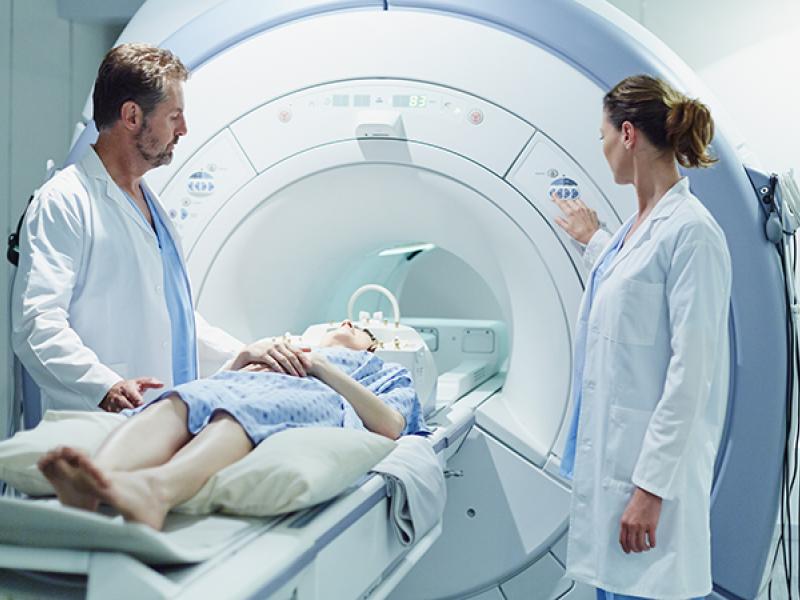
During this rotation, interpretation of nuclear studies including myocardial perfusion imaging, pharmacologic stress testing, first pass and gated radionuclide angiography will be performed. A basic knowledge of radiation safety, use of radiopharmaceuticals, and acquisition and processing of nuclear medicine images is gained. Fellows on this rotation are responsible for exercise treadmills. Fellows will be a part of the nuclear laboratory and work closely with the ordering providers as well as the laboratory staff. They will feel comfortable communicating study findings with the ordering providers when appropriate. They will be expected to interact respectfully with patients, families, and all members of the healthcare team.
Clinical training in Nuclear Cardiology involves working under a physician preceptor and mastery of the areas of learning defined by COCATS 4 Task Force 6.
There are two levels of training from which the fellow will select: level 1 or level 2. The fellows are expected to identify at the start of their training the level they plan to achieve.
Rotation leader
Dr. Thomas Dresser
COCATS level 1
This is the minimum requirement for the Cardiovascular Disease Fellowship. Time spent in the Nuclear Medicine clinic is a minimum of 2 months. Completion of procedures and skills of Table 1 and Table 2, COCATS 4 document is required (see below). A minimum of 100 cases of cardiac imaging is required. Normally 100 cases per month are performed. This training allows the Fellow to become conversant with the field of nuclear cardiology for application in general clinical management of cardiovascular patients.
COCATS level 2
This training is a minimum of 700 hours of clinic and didactic training. This requires 4 months in Nuclear Medicine clinic. Training includes 80 hours of didactic and clinic training in the basic science of radiopharmaceuticals, imaging equipment, and radiation safety. See Master Task List below. There are about 20 lectures with accompanying homework. There are two written exams. A minimum of 300 cases of cardiac imaging is required. This training qualifies the individual to become an Authorized User of radiopharmaceuticals, interpret studies, and they are eligible to take the board certifying exam in Nuclear Cardiology. The Fellow is expected to maintain their own training log (see attached) which will be verified by the preceptor.
Lectures
- Introduction to Nuclear Cardiology-Description of training requirement to be an Authorized User and overview of the basics of nuclear medicine myocardial perfusion imaging. By T. Dresser
- Basic Atomic Physics- Atomic structure, nuclides, table of elements and table of nuclides, types of radioactive decay, gamma rays, X-rays
- Charged particles and photon interactions-Ionization, Bremsstrahlung, Compton effect, photoelectric effect, stopping power, attenuation, half-value layer
- Radiation Detectors-Geiger counter, energy resolution, pulse height spectrum, FWHM, dead time, signal-to-voltage characteristics, ionization chamber, scintillators, photomultiplier tube
- Radiation Characteristics & Quantities, Counting Statistics-Radiation units, linear energy transfer, absorbed dose, dose equivalent, Quality Factor, effective dose, half-life (biologic, physical, effective) Frequency distribution, mean, variance, coefficient of variation, binomial distribution, Poisson distribution, Chi square
- Scintillation Camera and Collimators-Structure of gamma camera, pulse height analyzer, resolution, sensitivity, uniformity, linearity, collimator characteristics
- Image Quality: Compensation for Attenuation & Scatter-Resolution, scatter, sensitivity, contrast, signal-to-noise ratio, attenuation coefficient, energy windows.
- Radiopharmaceuticals, Stress methods for Myocardial Perfusion Imaging-Tl-201, Tc-agents, treadmill, adenosine, regadenoson, dipyridamole, dobutamine
- Camera performance standards-Floods, uniformity, special and energy resolution, phantoms
- SPECT reconstruction-Filtered backprojection
- Radionuclide Production and Radiopharmaceutical Formulation-radionuclides, reactor vs. accelerator production, types of equilibrium, radionuclide purity, Mo-99/Tc99m generator
- Cell Responses to Radiation-energy transfer of ionizing ration to cells, survival curves, relative biologic effect, dose rate, linear energy transfer, dosimetry, target organ vs. critical organ
- Radiation Safety-exposure limits, as low as reasonably achievable (ALARA), labeling of packages, posting of radiation areas.
- Image analysis, planar imaging-Windowing, brightness, contrast, artifacts; planar images
- PET imaging-Positron emission and cameras, Rb-82, F-18-FDG
- Risk stratification-Appropriate use criteria, incremental prognostic value of MPI, CARP trial
- Dynamic cardiac imaging-First pass, MUGA, gated images
- Acute chest pain evaluation-Protocols for evaluating acute chest pain in ED
- Artifacts-Attenuation, motion, extra-cardiac activity, MPI versus cath cases
Evaluation of learning
To receive full credit for training at Level II the Fellow will complete the homework assigned to each lecture in Table 1. There will be two written exams pertaining to the lectures and the Fellow is required to achieve a passing score of 80%. Each Fellow is responsible for recording their training on the “Nuclear Cardiology Training Log” listed below. Each item completed should be verified by the attending physician. The training log will be presented to the preceptor at completion of training.


